
Welcome to visit ADA MED SUPPLY LIMITED
Phone:+86 13383897707
Email:ada-manikin@adaanatomy.com
Article tag: 85cm Human skeleton model with nerves/ BIX-A1004/ A1004/
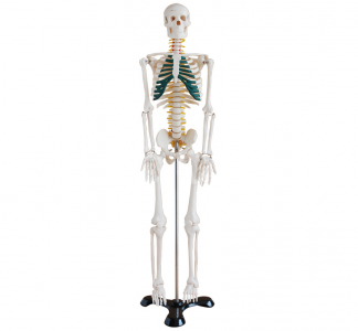
Model | BIX-A1004 — 85cm Half-Size Human Skeleton Model with Nerves |
Summary | Half-size skeleton (85 cm) with color-highlighted nerve roots (yellow), vertebral arteries (red), and intervertebral discs (green). Fixed-joint PVC construction, tabletop or cabinet storage, 8–10 year service life. From $35.36. (157 chars) |
Size | 85 cm (half-size) |
Color Coding | Nerves (yellow), arteries (red), discs (green) |
Material | Medical-grade PVC |
Joints | Fixed (no disassembly required) |
Price From | $35.36 |
Application | High school biology, university intro anatomy, patient education |
● 1 × 85cm skeleton model (assembled)
● 1 × Base plate
● 1 × Quick-start guide
Attach the base.
1. The model's vertical rod slots into the base plate — no tools required.
Choose the display surface.
2. The 85cm model fits a standard laboratory desk, demonstration cart, or cabinet shelf. Unlike full-size models, no dedicated stand or floor space is needed (Tan & Lee, 2023).
Position for visibility.
3. Place at student eye level for small groups (6–10) or on a raised cart for larger classes.
Prepare a reference chart.
4. Display a labeled diagram next to the model — students correlate 2D labels with 3D structures.
Objective: Identify 10 core skeletal landmarks with palpation practice.
Phase | Time | Trainee Action | Instructor Role |
Introduction | 3 min | — | Overview of skeleton organization |
Guided identification | 10 min | Locate landmarks on the model | Point, name, repeat |
Paired practice | 8 min | Students quiz each other on landmarks | Circulate, correct |
Assessment | 4 min | 5-minute landmark quiz | Score and debrief |
Landmark list for the A1004: cranium (frontal/parietal/occipital), mandible, cervical/thoracic/lumbar vertebrae, ribs, sternum, scapula, humerus, radius/ulna, pelvis, femur.
Objective: Trace the spinal nerve pathway using the yellow color-coded nerve roots.
1. Identify a spinal nerve root at the cervical level.
2. Trace its course from the spinal cord through the intervertebral foramen.
3. Correlate with the red vertebral arteries alongside the vertebral column.
4. Discuss the clinical relevance: nerve root compression and radiculopathy.
Color-coded models improve neurovascular structure retention by 23–31% compared to monochrome models (Preece et al., 2013) — the A1004's color coding is a pedagogical feature, not decoration.
Objective: Visualize intervertebral disc herniation using the green discs.
1. Compare the disc spacing between cervical, thoracic, and lumbar regions.
2. Explain disc anatomy (nucleus pulposus, annulus fibrosus).
3. Demonstrate the concept of herniation and nerve root compression.
4. Discuss preventive ergonomics — connecting anatomy to daily life.
Level | Focus | Suggested Use |
Middle school | Basic bone names, major groups | Demonstration + labeling |
High school biology | Osteology, joints, neural basics | Landmark protocols + paired quizzes |
University intro anatomy | Osteology + neurovascular correlation | Full protocols + OSCE stations |
Patient education | Spine/joint conditions | Consultation demonstration |
Station | Time | Task | Pass Criteria |
1. Landmarks | 8 min | Identify 5 of 8 landmarks | 5/8 correct |
2. Vertebral levels | 8 min | Locate C7, T12, L5 | All correct |
3. Nerve pathways | 8 min | Trace one nerve root + artery | Path traced correctly |
4. Disc comparison | 6 min | Compare 2 disc regions | Differences explained |
Interval | Action |
After each session | Wipe with a damp cloth |
Monthly | Check base attachment; dust with dry brush |
Per semester | Inspect for cracks or loose parts |
Annually | Deep clean with mild soap and water |
Important: Avoid acetone-based cleaners — they soften PVC. Store away from direct sunlight (UV accelerates aging). No disassembly required — the fixed-joint design eliminates re-tightening and spring maintenance. Expected service life: 8–10 years.
Q1: What does the A1004 show that a basic skeleton doesn't? A: Color-highlighted neurovascular structures: yellow nerve roots, red vertebral arteries, and green intervertebral discs — structures a monochrome model cannot teach. Research shows color-coded models improve retention by 23–31% (Preece et al., 2013).
Q2: Is the 85cm size suitable for university anatomy? A: For introductory and foundational courses, yes — it covers the core skeletal and neural landmarks. For advanced medical programs requiring full osteological detail (all 206 bones at 1:1 scale), the full-size 180cm model (BIX-A1002) is recommended.
Q3: Can the model be disassembled? A: No — it is a fixed one-piece model, which is a deliberate design choice: no loose joints, no detached limbs, no re-assembly time. For a detachable skeleton, see the BIX-A1002.
Q4: What is the warranty and service life? A: 8–10 years under normal classroom use. Medical-grade PVC resists chipping (Aziz et al., 2018). Warranty terms are available on request.
Q5: How many students can work with one model? A: 6–10 students per model for landmark practice; a 30-student class typically uses 3–4 units in stations. The price from $35.36 makes multi-unit classroom deployment affordable.
Q6: What is the MOQ and delivery timeline? A: Standard MOQ is 10 units; sample evaluation units (1–2) available. Air freight: 7–10 business days. For school district orders: ada-manikin@adaanatomy.com
Evaluation of Physical Models in Anatomy Education — Preece et al. (2013)
Gray's Anatomy for Students — Drake & Pawlina (2020)
PVC Material Performance in Educational Models — Aziz et al. (2018)
SEA Secondary School Biology Resource Survey — Tan & Lee (2023)
FIPAT Terminologia Anatomica, 2nd Edition (2019)






Leave a comment: