
Welcome to visit ADA MED SUPPLY LIMITED
Phone:+86 13383897707
Email:ada-manikin@adaanatomy.com
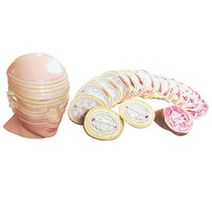

Students learn anatomy in layers and then meet it in slices. The BIX-A3001 closes that gap: 17 serial cross sections of the head and neck, each about 1.2 cm thick, stacked into a 19 × 17.5 × 24.5 cm model at a listed price of US$397.80.
| Item | Detail |
|---|---|
| Model | BIX-A3001 |
| Category | Anatomical series / head and neck |
| Sections | 17 |
| Section thickness | Approximately 1.2 cm |
| Size | 19 × 17.5 × 24.5 cm |
| Teaching focus | Cross-sectional anatomy, radiology orientation, sectional dissection revision |
| Listed price | US$397.80 |
| Certification | ISO 9001 / 14001 / 45001 and CE documentation on request |
A diagram of a single slice teaches one level. Seventeen stacked slices teach the third dimension: how the airway moves from nasopharynx to larynx, how the carotid sheath migrates laterally, how the parotid gland gives way to the masticator space. Those relationships are exactly what a CT or MRI reader reconstructs mentally, and they are almost impossible to teach from a single transverse image.
Three-dimensional visualisation has a documented learning advantage over flat images in anatomy education, with meta-analytic evidence favouring physical 3D models for spatial understanding [1]. Serial sections add sequence: the model is not one 3D object but 17 2D objects arranged in the order a scanner would acquire them.
Method matters as much as the object. A BEME systematic review of anatomy teaching found no single format that dominates, but concluded that combining didactic instruction with hands-on, spatially explicit tools produces better retention than lectures alone [3]. Deliberate practice with specific feedback is the mechanism most consistently identified behind that gain [2] — which is why the class plan below spends its time on retrieval and matching, not on a tour of the model.
1. Orientation before content. Ask students to find anterior, posterior, left and right on a single slice before naming any structure. Orientation errors cause most of the misidentification seen in early radiology teaching.
2. Airway continuity. Trace the nasal cavity, pharynx, larynx and trachea across consecutive slices. Students who can follow the airway rarely confuse the levels afterwards.
3. Vessel and nerve bundles. The carotid sheath, the internal jugular vein and the vagus sit together; following them across sections shows why a mass at one level displaces structures at the next.
4. Spaces and fascia. The parapharyngeal, retropharyngeal and submandibular spaces become visible as regions between structures rather than as named boxes on a diagram.
5. Radiology cross-check. Place a printed CT slice beside the matching model section. The exercise is simple and it is where the model earns its price.
Knowing these limits prevents the most common complaint after purchase — that the model "does not look like a real scan".
Q1. How many sections does the BIX-A3001 have? 17 serial cross sections of the head and neck.
Q2. How thick is each section? Approximately 1.2 cm, which is coarser than a clinical CT but suitable for teaching relationships.
Q3. What are the model dimensions? 19 × 17.5 × 24.5 cm assembled.
Q4. Is it suitable for radiology teaching? Yes for orientation and cross-sectional relationships; for slice-by-slice scrolling, pair it with imaging software.
Q5. What is the price and minimum order? The listed price is US$397.80; MOQ is flexible. Email for the current price list and volume tiers.
Q6. How do we request a quotation or sample? Email ada-manikin@adaanatomy.com with quantity, city and course level for pricing, packing data and lead time.
For the specification sheet, section list and a department quotation: ada-manikin@adaanatomy.com. Production follows ISO 45001 management practice; CE documentation is available for EU procurement.






Leave a comment: