
Welcome to visit ADA MED SUPPLY LIMITED
Phone:+86 13383897707
Email:ada-manikin@adaanatomy.com
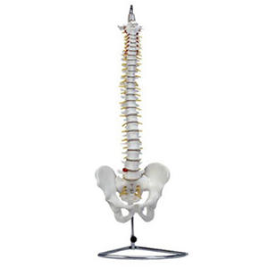
Students memorise vertebral counts quickly and then fail at the questions that matter clinically: which root exits at which level, where the vertebral artery runs, and why a lower lumbar disc herniation produces a specific dermatome. A life-size spine with pelvis answers those questions spatially. The BIX-A1014 does it at natural size — a 29-inch spine mounted on a 34-inch iron base, with nerve roots, vertebral arteries, discs, occiput and humerus included — for US $47.14 as listed.

| Item | Detail |
|---|---|
| Model | BIX-A1014 |
| Scale | Natural size |
| Spine height | 29 inches (≈74 cm), mounted on a 34-inch iron base |
| Structures shown | Vertebral column, nerve roots, vertebral arteries, intervertebral discs, pelvis, humerus, occipital bone, lumbar disc |
| Key features | Transverse processes and spinal incisions modelled for demonstration |
| Listed price | US $47.14 (confirm current price and freight) |
| Typical users | Chiropractic, orthopaedics, physiotherapy, anatomy departments, corporate health programmes |
At natural size, the model can be placed beside a standing student and compared directly. That comparison is where spatial learning happens: the student sees that the cervical spine occupies roughly a quarter of the column, that the lumbar vertebrae are visibly larger, and that the sacrum sits between the iliac crests at a height they can find on their own body.
Research on anatomy teaching with physical and printed models keeps returning to the same finding: three-dimensional handling improves understanding of spatial relationships in a way that two-dimensional images do not [1]. The spine is the strongest case for that claim, because almost every clinical question about it is a question about relative position — medial to, anterior to, passing through.
The vertebral artery. It is easy to draw and hard to place. On the A1014 the artery is modelled in relation to the cervical transverse processes, which is exactly the relationship that matters when a student is asked why cervical manipulation carries a vascular risk, or where a vertebral artery dissection can occur.
The nerve roots and their exit levels. Because the model shows nerve roots emerging along the column, the C5–C6 versus L4–L5 distinction becomes visible rather than memorised. Ask students to trace a root from the cord to the intervertebral foramen and name the level; the model makes the answer checkable by eye.
The disc and the transverse process. These are the landmarks for injection and for interpreting imaging. A model that includes the lumbar disc and the transverse processes lets an instructor point at the target while a student holds the model.
Drill 1 — Level identification under time pressure (10 min). The instructor points at a vertebra; students name the region and the number within 5 seconds. Rotate the model between rounds so students cannot use position memory.
Drill 2 — Root-to-dermatome mapping (15 min). Give each pair a symptom (numb great toe, weak wrist extension, pain in the lateral foot) and ask them to identify the likely level on the model, then justify it by tracing the root.
Drill 3 — Imaging correlation (20 min). Show a sagittal MRI slice and ask students to find the equivalent level on the model. Radiology-integrated anatomy teaching has measurable educational value in field studies of anatomy curricula [2], and the model is what allows the correlation to be physical rather than abstract.
It is a static anatomical model, not a motion simulator: it shows structure and relationships, not coupled movement. If your curriculum requires flexion–extension or rotation analysis, you need a dynamic or articulated spine model — the literature on 3D-printed dynamic cervical spine models describes that use case specifically [1].
It also does not reproduce pathological states. For disc herniation, stenosis or fracture teaching, pair the normal model with pathological specimens or imaging cases.
Q1. Is the BIX-A1014 life-size?
Yes — natural size, with a 29-inch spine mounted on a 34-inch iron base.
Q2. Which structures are included?
The vertebral column, nerve roots, vertebral arteries, intervertebral discs, pelvis, humerus and occipital bone, with transverse processes and spinal incisions modelled for demonstration.
Q3. What is the listed price?
US $47.14 per unit as listed on the product page; confirm the current price, packing and freight with the supplier.
Q4. Is it suitable for chiropractic teaching?
Yes — the listing positions it for chiropractic, orthopaedic and allied health programmes, and the modelled transverse processes support technique demonstration.
Q5. Does it articulate?
No. It is a static anatomical model; for movement analysis choose an articulated or dynamic spine model.
Q6. How do we request a sample or quotation?
Email ada-manikin@adaanatomy.com with the quantity, your city and whether you need the display base.
For the specification sheet, packing details and a landed-cost quotation: ada-manikin@adaanatomy.com. Production follows ISO 45001 management practice; CE documentation available for EU procurement.






Leave a comment: