
Welcome to visit ADA MED SUPPLY LIMITED
Phone:+86 13383897707
Email:ada-manikin@adaanatomy.com
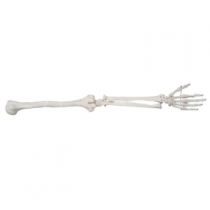
Short answer: BIX-A1031 is a life-size upper limb skeleton model in PVC, covering the humerus, radius, ulna and the bones of the hand. It is one item inside a series that also includes a life-size lower limb model and compact full-body skeletons — and the purchase decision is almost always between those, not between suppliers. Choose the life-size limb model when the teaching objective is the bone itself: its shape, its articular surfaces and its handling. Choose the limb version over a full skeleton when detail and pass-around convenience matter more than body-wide context.
Key takeaways
| Objective | Model to choose | Why |
|---|---|---|
| Teach the bones of the arm in detail | BIX-A1031 — life-size upper limb skeleton | Full-scale humerus, radius, ulna and hand bones; the model can be passed along a row. |
| Teach the bones of the leg in detail | BIX-A1032 — life-size lower limb skeleton | The equivalent life-size model for the lower extremity in the same series. |
| Give a quick body-wide overview | BIX-A1006 — 42 cm human skeleton | A compact study skeleton: the whole body at desk scale, without limb detail. |
| Combine skeleton with vessels or nerves | BIX-A1004 — 85 cm skeleton with nerves | Adds structures beyond bone, at reduced scale. |
| Study a single joint instead of a whole limb | Hand joint / foot joint joint-models in the same series | Narrower focus, smaller footprint, cheaper per station. |
| Published data — BIX-A1031 | Value on the product page |
|---|---|
| Product model | BIX-A1031 |
| Scale | Life size |
| Material | PVC |
| Structures | Upper limb skeleton: humerus, radius, ulna, hand bones |
| Certification | ISO and CE (as published) |
| Price | Contact us to obtain the price |
| Package dimensions / weight | Not published on the page |
A full skeleton answers "where is this?" A limb model answers "what is this, and how does it move?" They are different questions, and the second one is where students lose marks. The appendicular skeleton — the limbs — is the part of the skeleton that clinicians interpret most often on plain radiographs, and dedicated short training in appendicular skeletal image interpretation has been shown to change reporting accuracy [1].
The case for a physical model over a picture is also direct: in a randomised comparison of physical 3D-printed models against cadaveric material for learning anatomy, the printed physical models performed comparably [2]. If a printed model can carry that load, a purpose-made life-size PVC limb can certainly carry the teaching of its own bones — with the advantage that it is durable and can be handled by every student in the room.
Send these questions to ada-manikin@adaanatomy.com and we will answer each point and attach the current supply data.
Q1. What is BIX-A1031?
It is a life-size upper limb skeleton model made of PVC, used for teaching the bones of the arm.
Q2. Which bones does it include?
The published description names the humerus, radius, ulna and the bones of the hand.
Q3. What material is it made from?
PVC.
Q4. Should I buy this or a full-body skeleton?
Buy the limb model when the objective is detailed bone anatomy and handling; buy a compact full skeleton (such as the 42 cm model) when the objective is body-wide orientation.
Q5. Is it certified?
ISO and CE certification are published with the product. Request the certificate copies for procurement records.
Q6. How do I get the price and the matching lower limb model?
Email ada-manikin@adaanatomy.com with the model (BIX-A1031) and the quantity; ask for BIX-A1032 in the same message if you need the lower limb as well.
Write to ada-manikin@adaanatomy.com for a quotation on BIX-A1031 and its lower limb counterpart BIX-A1032. If your course also covers joints, ask about the hand and foot joint models in the same series — they extend the same teaching sequence at lower cost per station.






Leave a comment: