
Welcome to visit ADA MED SUPPLY LIMITED
Phone:+86 13383897707
Email:ada-manikin@adaanatomy.com
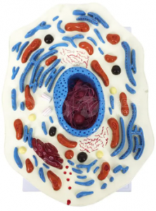
Short answer: The BMN/B054 is an animal cell model enlarged 20,000 times. It shows the cell membrane, the cytoplasm and the nucleus, and the cytoplasm is built out with the structures that matter most in an introductory course: mitochondria, rough and smooth endoplasmic reticulum, the Golgi complex and the centrosome. The cut surface reveals the nuclear membrane, the nucleolus and chromosomes, and the model carries digital indicators for the labelled parts.
Key takeaways
| Item | Specification on the product page |
|---|---|
| Magnification | 20,000× |
| Overall structures | Cell membrane, cytoplasm, nucleus |
| Cytoplasmic organelles | Mitochondria, rough endoplasmic reticulum, smooth endoplasmic reticulum, Golgi complex, centrosome |
| Cut-surface details | Nuclear membrane, nucleolus, chromosomes |
| Labelling | Digital indicators for multiple parts |
| Price | Inquiry for best offer |
| Certification | ISO and CE |
In a diagram, organelles are adjacent shapes. In a hand-held model, they have volume and position — the Golgi complex sits near the nucleus, the rough ER wraps around it, the mitochondria are distributed through the cytoplasm. That spatial knowledge is exactly what students need when they later learn what each structure does.
The educational case for three-dimensional cellular models is documented. A study of cellular models used in three dimensions reported improved learning of cell structure and function compared with conventional approaches [1]. The broader pattern holds for physical models in anatomy and biology teaching [2], and controlled comparisons have found three-dimensional physical models superior to two-dimensional computer presentations for this kind of learning [3].
There is also a practical argument. A biology lab that cannot afford electron micrographs of its own still needs students to know the difference between rough and smooth ER, and a durable classroom model delivers that knowledge every year.
Q1. What is the BMN/B054?
It is a model of an animal cell enlarged 20,000 times, showing the cell membrane, cytoplasm and nucleus together with the main organelles.
Q2. Which organelles are modelled?
Mitochondria, rough and smooth endoplasmic reticulum, the Golgi complex and the centrosome, plus the nuclear membrane, nucleolus and chromosomes at the cut surface.
Q3. Why is the magnification 20,000×?
At that scale the organelles become large enough to inspect and to label physically, which is the point of the model.
Q4. Can it be used for assessment?
Yes. The digital indicators allow structures to be identified by number, which works well for a quick oral or written quiz.
Q5. Is the model certified?
The product page states ISO and CE certification.
Q6. How do I get the best price?
Email ada-manikin@adaanatomy.com with the model (BMN/B054), the quantity you are considering and your destination port; the page quotes on request and our export team will reply with pricing, lead time and configuration.
Email ada-manikin@adaanatomy.com for the BMN/B054 best-offer price and shipping details. If your course covers cell division as well as cell structure, the same biology range includes a mitosis model and a meiosis model, so the genetics and cell-biology sections can be equipped in one order.






Leave a comment: