
Welcome to visit ADA MED SUPPLY LIMITED
Phone:+86 13383897707
Email:ada-manikin@adaanatomy.com
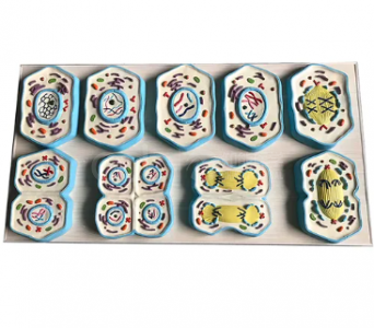
Short answer: The BMN/HB134 is an eight-part model of cell mitosis. It shows nuclear division from the first appearance of chromosomes in the nucleus through the five main stages of the process — interphase, prophase, metaphase, anaphase and the final separation into two daughter nuclei — and then cytokinesis, where a new cell wall forms between the two nuclei and one mother cell becomes two daughter cells. The nuclear membrane is modelled as completely transparent, so students can see the chromosomes inside each stage instead of having to imagine them.
Key takeaways
| Part / stage | What students see |
|---|---|
| Interphase | Chromosomes appear inside the nucleus before division begins |
| Prophase | Chromosomes condense; the nuclear membrane is still modelled as transparent |
| Metaphase | Chromosomes aligned at the cell equator |
| Anaphase | Chromatids separated and moving to opposite poles |
| Two daughter nuclei | Final state of nuclear division |
| Cytokinesis | New cell wall forming between the two new nuclei |
Cell division is a temporal process drawn in a static textbook, and that mismatch is where students lose the thread: the same chromosomes have to be tracked through five shapes, then a wall has to appear between two nuclei that did not exist at the start. The peer-reviewed literature on 3D cellular models supports exactly this point. A 2023 teaching study in the Journal of Microbiology & Biology Education notes that students struggle to visualise spatial relationships between cells and macromolecules, and reports that 3D printed custom cell models increased student learning about cell size and diffusion [1]. The reasoning transfers directly: when students can rotate the object in their hands, the spatial relationship stops being an act of imagination.
The broader evidence base for physical teaching models is unusually strong. Yammine and Violato's meta-analysis in Advances in Health Sciences Education (2016) pooled 8 studies — 7 of them randomised, with 16 comparison arms and 820 learners — and found that physical models as a low-cost three-dimensional method produced better factual and spatial knowledge outcomes than comparator teaching methods [2]. Their earlier meta-analysis in Anatomical Sciences Education (2015), covering 36 studies of three-dimensional visualisation in anatomy teaching, reached the same direction of effect and warned that many graduates leave with deficient structural knowledge [3].
The product page describes the eight-part composition and the five stages above, and lists a reference unit price of $117.86 that is explicitly subject to order quantity — so treat it as an indicative figure and request a quotation for a firm figure. Packaging and spare-part details are listed as a separate tab on the product page. This is a teaching model; it is not a laboratory specimen.
Q1. What is the BMN/HB134?
An eight-part teaching model of cell mitosis, covering nuclear division from interphase to two daughter nuclei plus cytokinesis.
Q2. How many stages does it show?
Five stages of nuclear division — interphase, prophase, metaphase, anaphase and the daughter nuclei — followed by cytokinesis and the formation of the new cell wall.
Q3. Can students see the chromosomes?
Yes. The nuclear membrane is modelled as completely transparent, so the chromosomes remain visible inside the nucleus at each stage.
Q4. What is the price?
The product page lists a reference unit price of $117.86, subject to the order quantity. Email ada-manikin@adaanatomy.com for a firm quotation.
Q5. Which level is it aimed at?
Secondary-school biology, college cytology and histology courses, and teacher-training demonstrations.
Q6. Can it be combined with a meiosis model?
Yes. Comparing the mitotic and meiotic sets side by side is one of the most effective ways to teach the difference between the two divisions; ask us for the Biology Series list.
Email ada-manikin@adaanatomy.com with the model (BMN/HB134), the quantity and the destination country. If your course covers the full Biology Series — cell models, mitosis and meiosis — we can quote the set together so a term's practicals are covered by one order.






Leave a comment: