Welcome to visit ADA MED SUPPLY LIMITED
Phone:+86 13383897707
Email:ada-manikin@adaanatomy.com
Article tag: eyeball and orbital magnification model/ BIX-A1062/
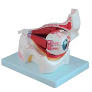
The eyeball and orbit magnification model, by proportionally magnifying the structure of the eyeball and the anatomical position of the orbit, enables the complex and tiny tissues to be clearly presented. It is a very important tool in ophthalmology teaching and training. Models typically display key structures such as the cornea, iris, lens, retina, and optic nerve, and present the directions of extraocular muscles, orbital blood vessels, and nerves, which helps learners understand the mechanism of vision formation and the spatial layout within the orbit.
In teaching, this model can help students quickly master the stratification of the eyeball, the path of light refraction and the functions of each part from a macro perspective. In clinical training, teachers can use models to explain the mechanism of ocular trauma, common lesion locations and related surgical approaches, thereby enhancing learners' spatial imagination ability and operational understanding.
In addition, it is also suitable for popular science exhibitions, enabling non-professionals to have an intuitive understanding of the structure of the eyes and the importance of vision protection. It is a commonly used efficient anatomy teaching auxiliary device in schools, hospitals, and medical training rooms.


Sophie Asveld
February 14, 2019
Email is a crucial channel in any marketing mix, and never has this been truer than for today’s entrepreneur. Curious what to say.
Sophie Asveld
February 14, 2019
Email is a crucial channel in any marketing mix, and never has this been truer than for today’s entrepreneur. Curious what to say.