Welcome to visit ADA MED SUPPLY LIMITED
Phone:+86 13383897707
Email:ada-manikin@adaanatomy.com
Article tag: Anatomical model of lower limb muscles/ BIX-A1102/
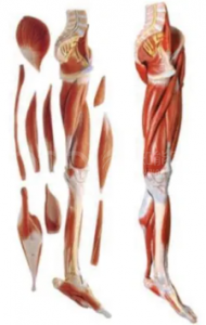
The lower limb muscle anatomy model is an intuitive teaching aid specifically designed for medical, nursing and sports rehabilitation education, used to demonstrate the structure, layers and functions of the lower limb muscles in the human body. This model is usually made to a real scale, clearly marking the positions and starting and end points of major muscle groups such as the quadriceps femoris, gastrocnemius, gluteus maximus, serratus, and biceps femoris. Some models can also be disassembled to facilitate the observation of the distribution of deep muscles and the course of nerves and blood vessels.
Through the anatomical model of lower limb muscles, learners can intuitively understand the working principles of lower limb muscles in movements such as walking, jumping, flexion and extension, and master the coordination relationships and anatomical characteristics among various muscle groups. In clinical teaching, this model helps students learn operational skills such as intramuscular injection, rehabilitation massage, and sports injury assessment, while assisting physical therapy and sports medicine professionals in analyzing muscle dysfunction and rehabilitation pathways.
The lower limb muscle anatomy model not only enhances the three-dimensional effect and practicality of anatomy teaching, but also effectively makes up for the limitations of traditional image teaching, enabling learners to master the knowledge and clinical application of lower limb anatomy more intuitively and accurately.


Sophie Asveld
February 14, 2019
Email is a crucial channel in any marketing mix, and never has this been truer than for today’s entrepreneur. Curious what to say.
Sophie Asveld
February 14, 2019
Email is a crucial channel in any marketing mix, and never has this been truer than for today’s entrepreneur. Curious what to say.