Welcome to visit ADA MED SUPPLY LIMITED
Phone:+86 13383897707
Email:ada-manikin@adaanatomy.com
Article tag: Foot joint model/ BIX-A1027/
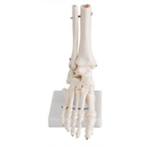
The foot joint model is a commonly used anatomical model in medical teaching and rehabilitation training. Its main function is to help learners intuitively understand the structure and movement characteristics of the ankle joint and foot joints.
This model is designed in proportion to the human body, truly reproducing the joint structure between the tibia, fibula and talus, calcaneus and metatarsal bones, and clearly showing the distribution of ligaments, joint capsules and some tendons. Through this model, trainees can systematically master the flexion, extension, varus and valgus movements of the ankle joint, and have an intuitive understanding of joint stability and the principles of motion mechanics. Some advanced models can also be disassembled and flexibly demonstrated, facilitating the observation of internal structures and clinical injury mechanisms, such as sprains, ligament strains, and pathological changes in arthritis.
This model is not only an important tool for teaching anatomy, sports medicine and rehabilitation in medical colleges and universities, but also applicable to clinical training and rehabilitation guidance, helping students and medical staff deepen their understanding and mastery of the ankle joint. Through intuitive demonstrations and repeated operations, learners' spatial cognition and clinical application abilities can be effectively enhanced, which is of great significance in the practice of prevention, treatment and rehabilitation of sports injuries.


Sophie Asveld
February 14, 2019
Email is a crucial channel in any marketing mix, and never has this been truer than for today’s entrepreneur. Curious what to say.
Sophie Asveld
February 14, 2019
Email is a crucial channel in any marketing mix, and never has this been truer than for today’s entrepreneur. Curious what to say.