Welcome to visit ADA MED SUPPLY LIMITED
Phone:+86 13383897707
Email:ada-manikin@adaanatomy.com
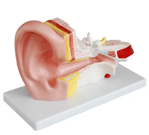
As an important tool in otolaryngology teaching, middle ear anatomical model provides an intuitive and efficient learning and training platform for medical students. The middle ear is a complex structure, containing tiny but functionally vital anatomical details such as the tympanic membrane, the auditory ossicle chain (malleus, incus, and stapes), and the eustachian tube. In practice, the diagnosis and treatment of middle ear related diseases are often accompanied by extremely high precision requirements, which poses a high challenge to the anatomical knowledge and operational skills of surgeons. So how can the anatomical model of the middle ear support this process?
1. Provide accurate anatomical learning support
The anatomical model of the middle ear can accurately reproduce the structure of the middle ear, especially the anatomical relationship between the auditory ossicle chain, the tympanic membrane and the surrounding tissue. Through three-dimensional display, this model enables students to clearly understand the anatomical layout and interrelationship of each part of the middle ear, especially the grasp of spatial position. For example, the connection between the malleus and the tympanic membrane, and the relationship between the stapes floor and the oval window, can be visually presented on the model, helping students to grasp the anatomical focus of the middle ear in a short time.

2. Simulate operation scenes to improve operation skills
Middle ear surgery is a highly delicate procedure involving complex technical requirements such as tympanic membrane repair and ossicular chain reconstruction. The middle ear anatomical model enables students to perform surgical simulations on the model by providing a realistic anatomical structure. For example, students can practice skills such as eardrum incision and ossicle manipulation without fear of harm to real patients. This low-risk training method helps to enhance the trainees' operational confidence and technical proficiency.
3. Enhance clinical judgment ability
In clinical ear surgery, accurate judgment of the lesion site is the key to successful treatment. For example, in surgery for chronic otitis media or otosclerosis, the surgeon needs to accurately identify the extent of damage to the ossicular chain and the location of the lesion. Middle ear anatomical model can simulate different pathological conditions (such as ossicular chain fracture, eustachian tube obstruction, etc.), so that students can improve their lesion recognition ability in training, so as to enhance their clinical judgment.
4. Data support the actual effect of the model
Studies have shown that students who use the middle ear anatomy model for training have significantly higher awareness of the middle ear anatomy structure and the success rate of surgical operations than students who do not use the model. According to the survey data of a medical training center, students who underwent model training in the simulation of myringotomy and stapes surgery improved the accuracy of the operation by about 30%. In addition, the simulation training also helped the trainees reduce the error rate in the actual operation.
conclusion
The middle ear anatomical model provides a strong support for the study and clinical training of medical students by providing intuitive anatomical display, simulated surgical scene and pathological situation. It not only improves students' understanding of middle ear anatomy and function, but also effectively enhances their operational skills and clinical judgment ability, ultimately laying a solid foundation for practical ear surgery.


Sophie Asveld
February 14, 2019
Email is a crucial channel in any marketing mix, and never has this been truer than for today’s entrepreneur. Curious what to say.
Sophie Asveld
February 14, 2019
Email is a crucial channel in any marketing mix, and never has this been truer than for today’s entrepreneur. Curious what to say.