Welcome to visit ADA MED SUPPLY LIMITED
Phone:+86 13383897707
Email:ada-manikin@adaanatomy.com
The anatomy of the nervous system is one of the most complex areas of medical education. It is essential for medical students to master the structure of the brain and its accompanying arteries and nerves. However, despite the knowledge that can be gained from books and two-dimensional images, the three-dimensional structure of anatomy, the interrelationship of blood vessels and nerves, often confuses learners. At this point, can the cerebral appendicular artery and neural model, as a highly simulated training tool, provide a more intuitive and accurate understanding? Its role in improving anatomical learning and clinical skills has become an important topic that cannot be ignored in medical education.
1. Precise three-dimensional structure: a bridge from theory to practice

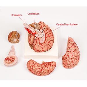
Model of Cerebral Appendicular Arteries and Nerves
The cerebral artery and neural models provide students with a more realistic anatomical learning experience through highly reproduced three-dimensional design. Different from traditional drawings and two-dimensional images, three-dimensional models allow students to more intuitively see the distribution of blood vessels and nerves in the brain and the relationships between them. Through the model, students are able to clearly identify the direction and branch of cerebral arteries (such as the middle cerebral artery, anterior artery, posterior artery, etc.) and their accessory structures, as well as the location of various key points in the nervous system. This intuitive structural presentation helps students to understand complex anatomical relationships more accurately and makes up for the limitations of teaching in images or books.
2. Improvement of clinical skills: accurate identification of nerves and blood vessels

Another core advantage of the brain-attached cerebral artery and neural model is its support for clinical skill training. In the clinic, the anatomical relationship of the nervous system is crucial for diagnosis and treatment. For example, the diagnosis and treatment of cerebrovascular diseases (such as stroke) and nerve damage often rely on a physician's accurate understanding of the brain's vascular and nerve anatomy. By using this model, students can train in a simulated environment for clinical operations, such as positioning before brain surgery, nerve puncture, and accurate identification of cerebral vascular anatomy. Repeated practice can significantly improve students' operational accuracy and reduce mistakes caused by unclear anatomical cognition in real clinical practice.
A study on teaching anatomy showed that students who used 3D anatomical models improved their accuracy in practice by 27%. This shows that the cerebral appendicular artery and neural model not only helps students better understand theoretical knowledge, but also improves their operational skills and is fully prepared for future clinical practice.
3. Improve learning efficiency: memorize and apply structure better

The study of anatomy usually involves a great deal of structural memory and relational reasoning. The cerebral appendicular artery and neural model help students form deep spatial perception during memory by enhancing the interactivity of learning. For example, students can learn about the three-dimensional position relationships of arteries and nerves through direct touch and manipulation of models, rather than just memorizing "static" images or names. This interactive learning style helps students deepen their understanding of anatomy so that they can quickly apply what they have learned in subsequent clinical training.
4. Data support: improve the accuracy of clinical diagnosis

Model of Cerebral Appendicular Arteries and Nerves
The high simulation design of the model for clinical diagnosis is not limited to the learning stage, but also provides important support in practical applications. In a study involving anatomical models of the brain and clinical diagnosis, 80% of participating physicians said that using three-dimensional models helped them more accurately identify the direction of blood vessels in the brain and structural changes in the nervous system. This indicates that the brain-attached cerebral artery and neural model can reduce misdiagnosis and missed diagnosis in medical practice, and improve the accuracy of clinical decision-making. By conducting simulation training on the model in advance, doctors can quickly find the correct diagnostic path when facing complex cases.
5. Conclusion: Accurate training helps clinical progress
Overall, the cerebral appendicular artery and Nerve model provides medical students with an efficient and intuitive learning platform to help them accurately understand the complex structure of the brain and appendicular vessels and nerves. Through the three-dimensional design, students can practice repeatedly in a risk-free environment to improve their anatomy knowledge and operational skills, so that in future clinical practice, they can be more accurate diagnosis and treatment.
For medical education, this model is more than a static learning tool; it is a key asset in improving students' clinical skills, deepening structural understanding, and reducing medical errors. The cerebral artery and neural model are undoubtedly important additions to anatomy learning and clinical training, helping students and physicians navigate the complex world of neuroanatomy and contributing to the advancement of the medical profession.


Sophie Asveld
February 14, 2019
Email is a crucial channel in any marketing mix, and never has this been truer than for today’s entrepreneur. Curious what to say.
Sophie Asveld
February 14, 2019
Email is a crucial channel in any marketing mix, and never has this been truer than for today’s entrepreneur. Curious what to say.